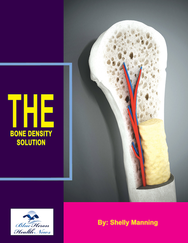
The Bone Density Solution by Shelly ManningThe program is all about healthy food and healthy habits. As we discussed earlier, we develop osteoporosis due to low bone density. Therefore, you will have to choose the right food to help your calcium and other vitamin deficiencies. In addition to healthy food, you will have to regularly practice some mild exercises. Your doctor might offer you the same suggestion. However, the difference is that The Bone Density Solution will help you with an in-depth guide.
Bone Density in Different Bones of the Body
Bone density, also known as bone mineral density (BMD), varies significantly across different bones in the body. These variations are influenced by factors such as the bone’s location, function, structure, and the type of bone tissue it contains—cortical or trabecular. Understanding the differences in bone density across various bones helps in assessing fracture risk, diagnosing conditions like osteoporosis, and developing targeted treatment strategies.
1. Long Bones: Femur, Tibia, and Humerus
Long bones are primarily composed of cortical bone in their shafts (diaphysis) and trabecular bone at their ends (epiphyses). These bones support body weight and facilitate movement, making them crucial for locomotion and load-bearing activities.
Femur (Thigh Bone):
- Bone Density: The femur has a high density in its cortical shaft, providing strength and resistance to bending. The femoral neck, which connects the shaft to the hip joint, contains more trabecular bone and is more susceptible to osteoporosis and fractures, particularly in older adults.
- Fracture Risk: Hip fractures are common in the elderly, often occurring at the femoral neck due to its lower density and the high metabolic activity of trabecular bone.
Tibia (Shin Bone):
- Bone Density: The tibia is also primarily composed of dense cortical bone, especially in the shaft. The density of the tibia is critical for weight-bearing and stability.
- Fracture Risk: Stress fractures can occur in the tibia due to repetitive load-bearing activities, particularly in athletes. These fractures are more likely when bone density is compromised by factors like osteoporosis.
Humerus (Upper Arm Bone):
- Bone Density: The humerus has a dense cortical shaft and a trabecular-rich head that connects to the shoulder. Bone density in the humerus is important for supporting arm movements and bearing loads.
- Fracture Risk: Fractures of the humeral head or shaft can occur due to falls or direct trauma, with the risk increasing in individuals with low bone density.
2. Spine: Vertebrae
Vertebrae are the bones that make up the spinal column. They are composed of a thin outer layer of cortical bone and a large amount of trabecular bone inside, which makes them particularly vulnerable to bone density loss.
Bone Density:
- Vertebrae have lower overall bone density compared to long bones due to the high proportion of trabecular bone. This makes the spine more susceptible to osteoporosis-related fractures.
- Age-Related Changes: As individuals age, the density of trabecular bone in the vertebrae decreases more rapidly than in cortical bone, leading to an increased risk of vertebral compression fractures.
Fracture Risk:
- Vertebral fractures are a common consequence of osteoporosis. They can lead to significant pain, loss of height, and kyphosis (a forward curvature of the spine).
3. Pelvis: Ilium, Ischium, and Pubis
The pelvis is a complex structure composed of several bones, including the ilium, ischium, and pubis. It supports the weight of the upper body and protects vital organs.
Bone Density:
- The pelvis has a mix of cortical and trabecular bone, with the iliac crest (the top part of the ilium) being rich in trabecular bone. The density of the pelvic bones is crucial for weight-bearing and stability.
- Age-Related Changes: Like the spine, the pelvis is vulnerable to osteoporosis, particularly in areas rich in trabecular bone.
Fracture Risk:
- Pelvic fractures are common in the elderly and can be particularly debilitating due to the pelvis’s central role in movement and weight-bearing.
4. Ribs: Costal Bones
Ribs are curved bones that form the rib cage, protecting the thoracic organs such as the heart and lungs. They are primarily composed of cortical bone but also contain trabecular bone in their inner layers.
Bone Density:
- Ribs have a moderate bone density, with a balance between cortical and trabecular bone. Their density is important for providing the rigidity needed to protect vital organs.
- Age-Related Changes: Bone density in the ribs decreases with age, making them more prone to fractures.
Fracture Risk:
- Rib fractures can occur from trauma, such as falls or impacts. In individuals with low bone density, fractures can occur even from minor impacts or severe coughing.
5. Skull: Cranial Bones
The skull is composed of several bones that protect the brain and form the structure of the face. It is primarily composed of cortical bone, with some trabecular bone in certain areas.
Bone Density:
- The cranial bones have a high density due to their thick cortical layer, which provides protection for the brain.
- Age-Related Changes: Bone density in the skull generally decreases with age, but fractures are less common due to the skull’s robust structure.
Fracture Risk:
- Skull fractures typically result from significant trauma, such as falls or direct impacts. While not as prone to osteoporosis-related fractures, the skull’s bone density is still important for overall cranial protection.
6. Forearm: Radius and Ulna
Forearm bones include the radius and ulna, which support the arm’s movement and stability. These bones are composed of both cortical and trabecular bone.
Bone Density:
- The radius and ulna have dense cortical shafts, with trabecular bone present near the joints, particularly at the wrist.
- Age-Related Changes: Bone density in the forearm can decrease with age, especially in the trabecular regions, leading to an increased risk of fractures.
Fracture Risk:
- Fractures of the radius, particularly at the wrist (Colles’ fracture), are common in individuals with osteoporosis. These fractures often occur due to falls.
Conclusion
Bone density varies significantly across different bones in the body, reflecting their unique functions and structural compositions. Long bones like the femur and tibia have high-density cortical shafts that provide strength and support, while the spine and pelvis, with their higher trabecular content, are more susceptible to density loss and fractures. Understanding these variations is crucial for assessing fracture risk, especially in conditions like osteoporosis, and for developing strategies to maintain bone health throughout the body.
The Bone Density Solution by Shelly ManningThe program is all about healthy food and healthy habits. As we discussed earlier, we develop osteoporosis due to low bone density. Therefore, you will have to choose the right food to help your calcium and other vitamin deficiencies. In addition to healthy food, you will have to regularly practice some mild exercises. Your doctor might offer you the same suggestion. However, the difference is that The Bone Density Solution will help you with an in-depth guide.
