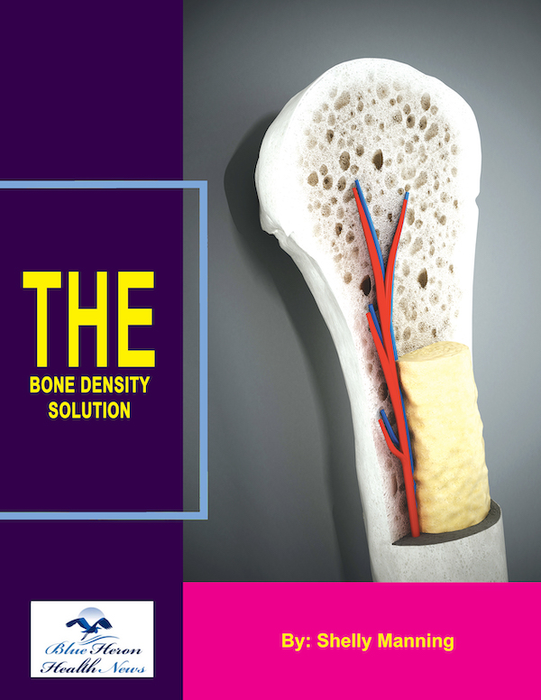
The Bone Density Solution by Shelly ManningThe program is all about healthy food and healthy habits. As we discussed earlier, we develop osteoporosis due to low bone density. Therefore, you will have to choose the right food to help your calcium and other vitamin deficiencies. In addition to healthy food, you will have to regularly practice some mild exercises. Your doctor might offer you the same suggestion. However, the difference is that The Bone Density Solution will help you with an in-depth guide.
Advances in Bone Density Measurement Techniques
Advances in Bone Density Measurement Techniques
Bone density measurement is a crucial tool in diagnosing osteoporosis, assessing fracture risk, and monitoring bone health over time. While Dual-Energy X-ray Absorptiometry (DEXA) has long been the gold standard, recent advances in technology have introduced new techniques and improvements to existing methods, offering more comprehensive insights into bone health. Here are some of the most significant advances in bone density measurement techniques.
1. High-Resolution Peripheral Quantitative Computed Tomography (HR-pQCT)
What It Is:
- HR-pQCT is an advanced imaging technique that provides high-resolution, three-dimensional images of bone structure, typically focusing on peripheral sites like the wrist and ankle.
Key Features:
- 3D Imaging: Unlike DEXA, which provides two-dimensional areal bone density, HR-pQCT offers detailed 3D images that allow for the separate analysis of cortical (outer layer) and trabecular (inner, spongy layer) bone.
- Bone Microarchitecture: HR-pQCT can assess bone microarchitecture, providing insights into bone quality, such as trabecular thickness, number, and spacing, which are crucial for understanding bone strength.
- Volumetric Bone Density: This technique measures true volumetric bone density, offering more precise data than the areal bone density provided by DEXA.
Applications:
- Research and Clinical Settings: HR-pQCT is particularly useful in research and specialized clinical settings where detailed bone quality analysis is needed. It is valuable in studying diseases that affect bone microarchitecture, such as osteoporosis and rheumatoid arthritis.
Limitations:
- Accessibility and Cost: HR-pQCT is more expensive and less widely available than DEXA, limiting its use primarily to research institutions and specialized clinics.
2. Trabecular Bone Score (TBS)
What It Is:
- TBS is a software-based analytical tool that enhances standard DEXA scans by assessing the texture of the bone image to provide information about bone microarchitecture.
Key Features:
- Bone Quality Assessment: TBS evaluates the trabecular structure of bone, which is not directly visible on a DEXA scan. It provides an indirect measure of bone quality and can help predict fracture risk independently of bone mineral density (BMD).
- Complementary to DEXA: TBS is often used in conjunction with DEXA scans to offer a more comprehensive assessment of bone health, particularly in patients with conditions like diabetes or those taking corticosteroids, where bone quality may be compromised despite normal BMD.
Applications:
- Fracture Risk Assessment: TBS is used to enhance fracture risk prediction, particularly in patients who may have normal or mildly reduced BMD but poor bone quality.
- Clinical Use: TBS is increasingly integrated into clinical practice to provide a more complete picture of fracture risk, especially in patients with borderline or atypical BMD results.
Limitations:
- Dependent on DEXA: TBS is not a standalone measurement; it requires a DEXA scan for its analysis.
3. Quantitative Ultrasound (QUS)
What It Is:
- QUS is a non-invasive technique that uses sound waves to assess bone density and quality, typically at peripheral sites such as the heel (calcaneus).
Key Features:
- No Radiation: QUS does not involve ionizing radiation, making it a safer option for frequent monitoring and for use in populations where radiation exposure is a concern (e.g., pregnant women, children).
- Assessment of Bone Quality: QUS provides information on bone elasticity and strength, offering insights into bone quality that go beyond simple density measurements.
- Portable and Cost-Effective: QUS devices are portable and less expensive than DEXA machines, making them more accessible in various healthcare settings.
Applications:
- Screening Tool: QUS is widely used for osteoporosis screening, particularly in primary care and community health settings where DEXA may not be available.
- Monitoring Bone Health: It can be used to monitor changes in bone density over time, especially in populations at risk for osteoporosis.
Limitations:
- Less Precision: QUS is generally less precise than DEXA, particularly for central skeletal sites like the spine and hip.
- Site-Specific: QUS typically measures bone density at peripheral sites, which may not fully reflect overall fracture risk.
4. Magnetic Resonance Imaging (MRI)
What It Is:
- MRI uses strong magnetic fields and radio waves to create detailed images of bone and soft tissue, providing insights into bone structure and quality.
Key Features:
- No Radiation: MRI does not expose patients to radiation, making it a safe alternative for assessing bone health, particularly in populations where radiation exposure is a concern.
- Bone Quality and Structure: MRI can assess bone quality, including bone marrow composition, trabecular bone structure, and the presence of bone microdamage, which are critical for understanding fracture risk.
- Whole-Body Imaging: MRI can provide comprehensive imaging of multiple skeletal sites, offering a broader view of bone health than site-specific measurements like DEXA.
Applications:
- Research and Complex Cases: MRI is used in research settings and for complex cases where detailed bone quality assessment is needed. It is particularly valuable in diagnosing conditions like osteonecrosis or in evaluating bone integrity after fractures.
Limitations:
- Cost and Accessibility: MRI is more expensive and less accessible than other bone density measurement techniques, limiting its routine use.
- Time-Consuming: MRI scans take longer to perform compared to other imaging modalities, which can be a drawback in clinical settings.
5. Dual-Energy X-ray Absorptiometry (DEXA) Improvements
What It Is:
- DEXA remains the gold standard for measuring bone mineral density, but recent advancements have improved its accuracy and applicability.
Key Features:
- Improved Resolution: Advances in DEXA technology have improved image resolution, allowing for more precise measurements of bone density and better differentiation between bone and soft tissue.
- Whole-Body Composition Analysis: Modern DEXA machines can also assess whole-body composition, including fat and lean muscle mass, providing a more comprehensive view of overall health.
- Enhanced Software: New software tools enhance DEXA’s capabilities, such as incorporating TBS for better bone quality assessment and improved fracture risk prediction algorithms.
Applications:
- Routine Clinical Use: DEXA continues to be widely used for routine osteoporosis screening, diagnosis, and monitoring.
- Body Composition Studies: The ability to measure body composition alongside bone density makes DEXA valuable in studies related to obesity, sarcopenia, and metabolic health.
Limitations:
- Radiation Exposure: Although DEXA uses low levels of radiation, it still involves exposure, which may be a concern for some patients.
- Cost and Availability: While DEXA is widely available, it is still more expensive than simpler techniques like QUS, making it less accessible in some settings.
6. Single-Photon Absorptiometry (SPA) and Dual-Photon Absorptiometry (DPA)
What They Are:
- SPA and DPA are older techniques that use a single or dual photon source to measure bone density, typically at peripheral sites like the forearm.
Key Features:
- Less Common: These techniques have largely been replaced by more advanced methods like DEXA and QUS due to their limitations in precision and applicability.
- Peripheral Measurement: SPA and DPA primarily assess bone density at peripheral sites, providing less comprehensive data on overall bone health.
Applications:
- Historical Use: SPA and DPA were once used in clinical practice but are now mostly of historical interest or in specific research settings.
Limitations:
- Outdated Technology: These methods are less precise and have largely been superseded by newer technologies like DEXA and HR-pQCT.
Conclusion
Advances in bone density measurement techniques have expanded our ability to assess bone health beyond simple density measurements, offering deeper insights into bone quality, structure, and overall fracture risk. While DEXA remains the standard for bone density testing, new technologies like HR-pQCT, TBS, and improved DEXA software have enhanced our understanding of bone health, allowing for more accurate diagnosis and personalized treatment strategies. These innovations are particularly valuable in research and specialized clinical settings, where detailed bone assessments are critical for managing complex cases of osteoporosis and other bone-related conditions.
The Bone Density Solution by Shelly ManningThe program is all about healthy food and healthy habits. As we discussed earlier, we develop osteoporosis due to low bone density. Therefore, you will have to choose the right food to help your calcium and other vitamin deficiencies. In addition to healthy food, you will have to regularly practice some mild exercises. Your doctor might offer you the same suggestion. However, the difference is that The Bone Density Solution will help you with an in-depth guide.
