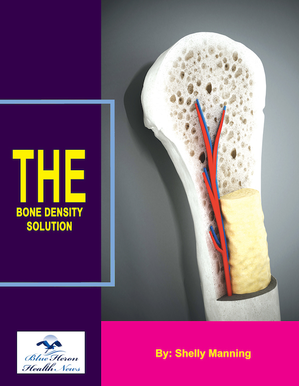
The Bone Density Solution by Shelly ManningThe program is all about healthy food and healthy habits. As we discussed earlier, we develop osteoporosis due to low bone density. Therefore, you will have to choose the right food to help your calcium and other vitamin deficiencies. In addition to healthy food, you will have to regularly practice some mild exercises. Your doctor might offer you the same suggestion. However, the difference is that The Bone Density Solution will help you with an in-depth guide.
How Osteoporosis is Diagnosed
Osteoporosis is a condition where bones become weak and brittle, leading to an increased risk of fractures. Because osteoporosis typically develops without obvious symptoms until a fracture occurs, it is often referred to as a “silent disease.” Early detection is crucial for managing the condition and preventing fractures. Here’s an overview of how osteoporosis is diagnosed:
1. Bone Mineral Density (BMD) Testing
The primary method for diagnosing osteoporosis is bone mineral density (BMD) testing, which measures the amount of minerals (such as calcium) in a specific area of bone. Lower bone density indicates an increased risk of fractures.
a. Dual-Energy X-ray Absorptiometry (DEXA or DXA) Scan
- Most common test: The DEXA scan is the gold standard for measuring bone mineral density and is used to diagnose osteoporosis.
- How it works: This test uses low-dose x-rays to measure the amount of mineral in your bones, typically in the spine, hip, or wrist. These areas are more likely to suffer fractures in people with osteoporosis.
- T-Score: The results are typically reported as a T-score, which compares your bone density with that of a healthy 30-year-old of the same sex.
- Normal: T-score of -1.0 or higher.
- Osteopenia (low bone mass): T-score between -1.0 and -2.5.
- Osteoporosis: T-score of -2.5 or lower.
- A T-score of -2.5 or lower indicates osteoporosis, and a score of -1.0 to -2.5 suggests osteopenia (a condition where bone density is low but not low enough to be classified as osteoporosis).
b. Quantitative Computed Tomography (QCT)
- Alternative to DEXA: This technique uses a CT scan to measure bone density, especially in the spine. It provides a 3D image and can assess bone quality and density in more detail than a standard DEXA scan.
- Disadvantage: It is more expensive and has a higher radiation dose than DEXA, so it’s typically not the first choice for routine screening.
2. Fracture Risk Assessment
For individuals who may be at risk but have not had a fracture, doctors often use tools that calculate the risk of fractures based on several factors. These tools can help assess the need for a bone density test and guide treatment decisions.
a. FRAX® (Fracture Risk Assessment Tool)
- What it is: FRAX is a computerized tool developed by the World Health Organization (WHO) that estimates the 10-year risk of having a hip or major osteoporotic fracture based on clinical risk factors.
- Risk factors used in FRAX:
- Age
- Gender
- Weight and height
- Previous fractures
- Family history of osteoporosis or fractures
- Smoking status
- Alcohol consumption
- Use of medications that affect bone health (e.g., corticosteroids)
- History of rheumatoid arthritis or other health conditions that increase fracture risk
- FRAX and BMD: It can also incorporate the BMD (T-score) if available, but it can also be used without this information.
3. Medical History and Physical Examination
Before conducting tests like a DEXA scan, a doctor will usually start with a medical history and physical examination.
- Medical History: The doctor will ask about risk factors such as:
- Age: Osteoporosis is more common in older adults.
- Family history: A family history of osteoporosis or fractures can increase the likelihood of the condition.
- Menstrual history: In women, menopause and age at first menstruation can influence bone density.
- Lifestyle: Smoking, alcohol use, and physical activity levels.
- Previous fractures: Even minor falls or injuries in the past can indicate weakened bones.
- Physical Examination: The doctor may check for signs that might suggest osteoporosis, such as:
- Height loss: People with osteoporosis often experience a reduction in height over time, as vertebrae in the spine become compressed.
- Posture: A stooped or hunched posture (kyphosis) may be a sign of spinal fractures due to osteoporosis.
- Fractures: Previous fractures, especially in the spine, hip, or wrist, could suggest the presence of osteoporosis.
4. X-rays
X-rays are not typically used to diagnose osteoporosis directly but can be helpful in certain situations:
- Fractures: If you have had a fracture or injury, an X-ray may show bone loss or fractures that could suggest osteoporosis.
- Spinal Compression: X-rays can detect compression fractures of the spine, which may be a result of osteoporosis.
However, bone density loss needs to be quite advanced before it shows up on regular X-rays. Therefore, DEXA scans are preferred for early detection.
5. Other Laboratory Tests (Blood and Urine Tests)
While blood and urine tests are not used to directly diagnose osteoporosis, they can help identify underlying conditions that may contribute to bone loss or rule out other potential causes of bone weakness.
- Blood tests: These can help assess calcium, vitamin D, and parathyroid hormone (PTH) levels, which are important for bone health. Abnormal levels could suggest an underlying condition affecting bone density.
- Calcium and Vitamin D: Low levels may indicate nutritional deficiencies that affect bone health.
- Parathyroid hormone (PTH): Elevated PTH levels can indicate issues with calcium regulation and bone resorption.
- Thyroid function tests: Thyroid disorders, like hyperthyroidism, can contribute to bone loss.
- Urine tests: Sometimes, doctors may test for urinary calcium and collagen breakdown products, which can help assess bone turnover and the rate of bone resorption.
6. Bone Biopsy
In rare cases, if there are concerns about other conditions affecting the bones, a bone biopsy might be performed. This involves removing a small sample of bone tissue to examine the structure under a microscope. This test is rarely used to diagnose osteoporosis but may be considered in certain situations to rule out other bone disorders, such as osteomalacia (softening of the bones) or Paget’s disease.
Conclusion
Osteoporosis is primarily diagnosed through bone mineral density (BMD) testing, most commonly using a DEXA scan, which helps measure bone density and assess fracture risk. Other methods, such as the FRAX® tool, medical history, physical exams, and blood tests, can also help provide a more comprehensive picture of an individual’s bone health. Early detection through regular screening and preventive measures can reduce the risk of fractures and improve outcomes for individuals at risk of osteoporosis.
The Bone Density Solution by Shelly ManningThe program is all about healthy food and healthy habits. As we discussed earlier, we develop osteoporosis due to low bone density. Therefore, you will have to choose the right food to help your calcium and other vitamin deficiencies. In addition to healthy food, you will have to regularly practice some mild exercises. Your doctor might offer you the same suggestion. However, the
