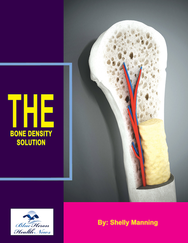
The Bone Density Solution by Shelly ManningThe program is all about healthy food and healthy habits. As we discussed earlier, we develop osteoporosis due to low bone density. Therefore, you will have to choose the right food to help your calcium and other vitamin deficiencies. In addition to healthy food, you will have to regularly practice some mild exercises. Your doctor might offer you the same suggestion. However, the difference is that The Bone Density Solution will help you with an in-depth guide.
How is the severity of low bone density assessed in India?
Assessing the severity of low bone density in India involves a comprehensive approach that includes clinical evaluation, bone density testing, risk factor assessment, and consideration of any existing fractures or symptoms. The following methods and criteria are used to evaluate the severity of low bone density:
Bone Density Testing
- Dual-Energy X-ray Absorptiometry (DEXA or DXA)
- Description: DEXA is the most widely used and preferred method for measuring bone mineral density (BMD). It uses low-dose X-rays to scan the bones, typically focusing on the hip and spine, which are common sites for osteoporotic fractures.
- Procedure: The patient lies on a padded table while the DEXA machine passes over the body. The machine measures the amount of X-rays absorbed by the bones, providing detailed images and data on bone density.
- Results: BMD results are expressed as T-scores and Z-scores:
- T-score: Compares the patient’s BMD to a young healthy adult’s BMD.
- Normal: T-score of -1.0 and above
- Osteopenia: T-score between -1.0 and -2.5
- Osteoporosis: T-score of -2.5 and below
- Z-score: Compares the patient’s BMD to others of the same age, sex, and body size. A Z-score of -2.0 or lower suggests a level of bone density that is significantly below the expected range for the patient’s age (Nature) (World Health Organization (WHO)) (IHCI).
- T-score: Compares the patient’s BMD to a young healthy adult’s BMD.
- Quantitative Computed Tomography (QCT)
- Description: QCT uses CT scans to provide three-dimensional images of the bones, allowing for separate measurements of cortical and trabecular bone density.
- Procedure: A CT scanner takes cross-sectional images of the bones, which are analyzed to determine bone density. QCT is particularly useful for evaluating spinal bone density.
- Usage: Less common than DEXA, QCT is available in specialized centers and is used when detailed bone architecture information is needed (World Health Organization (WHO)).
- Peripheral Devices
- Peripheral Dual-Energy X-ray Absorptiometry (pDXA): Measures bone density at peripheral sites such as the wrist or heel. It is useful for initial screening but less comprehensive than central DXA.
- Quantitative Ultrasound (QUS): Uses sound waves to measure bone density, typically at the heel. It is a quick and portable method but not as precise as DEXA (Nature).
Clinical Evaluation and Risk Assessment
- Clinical History and Physical Examination
- Medical History: Includes a detailed assessment of the patient’s family history of osteoporosis, personal history of fractures, and other risk factors such as hormonal imbalances, chronic diseases, and medication use.
- Physical Examination: Checks for signs of fractures, height loss, spinal deformities, and general bone health (World Health Organization (WHO)) (IHCI).
- Risk Factor Assessment
- FRAX Tool: The Fracture Risk Assessment Tool (FRAX) is used to estimate the 10-year probability of fractures in individuals. It incorporates clinical risk factors such as age, gender, body mass index (BMI), previous fractures, parental history of hip fracture, glucocorticoid use, rheumatoid arthritis, secondary osteoporosis, smoking, and alcohol use, along with BMD measurements.
- Lifestyle and Dietary Assessment: Evaluates the patient’s diet, physical activity levels, smoking status, and alcohol consumption, which are important factors influencing bone health (Nature) (World Health Organization (WHO)).
Laboratory Tests
- Biochemical Markers
- Serum Calcium: Measures the level of calcium in the blood, which is crucial for bone health.
- Vitamin D Levels: Assesses vitamin D status, which is essential for calcium absorption.
- Parathyroid Hormone (PTH): Evaluates the function of the parathyroid glands, which regulate calcium levels.
- Bone Turnover Markers: Includes tests for markers of bone formation (e.g., alkaline phosphatase) and bone resorption (e.g., C-terminal telopeptide) (World Health Organization (WHO)) (IHCI).
Imaging Studies
- Spinal X-rays
- Description: X-rays of the spine can detect vertebral fractures, which are common in osteoporosis and indicate severe bone loss.
- Usage: Useful for diagnosing fractures and deformities that result from low bone density (IHCI).
Conclusion
The severity of low bone density in India is assessed through a combination of bone density tests (primarily DEXA scans), clinical evaluations, risk factor assessments, laboratory tests, and imaging studies. Early detection and assessment are crucial for effective management and prevention of fractures. Increasing awareness and accessibility of these diagnostic tools are vital for improving bone health outcomes in India (Nature) (World Health Organization (WHO)) (IHCI).
References
- Mayo Clinic – Bone Density Test
- National Osteoporosis Foundation
- International Osteoporosis Foundation
- Journal of Clinical Densitometry
The Bone Density Solution by Shelly ManningThe program is all about healthy food and healthy habits. As we discussed earlier, we develop osteoporosis due to low bone density. Therefore, you will have to choose the right food to help your calcium and other vitamin deficiencies. In addition to healthy food, you will have to regularly practice some mild exercises. Your doctor might offer you the same suggestion. However, the difference is that The Bone Density Solution will help you with an in-depth guide.
