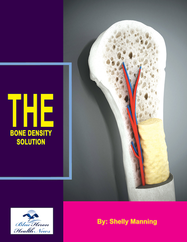
The Bone Density Solution by Shelly ManningThe program is all about healthy food and healthy habits. As we discussed earlier, we develop osteoporosis due to low bone density. Therefore, you will have to choose the right food to help your calcium and other vitamin deficiencies. In addition to healthy food, you will have to regularly practice some mild exercises. Your doctor might offer you the same suggestion. However, the difference is that The Bone Density Solution will help you with an in-depth guide.
How Bone Density is Measured
Bone density, or bone mineral density (BMD), is measured using various techniques designed to assess the amount of mineral content, particularly calcium, within a specific volume of bone. The most common and widely used methods for measuring bone density include Dual-energy X-ray Absorptiometry (DEXA or DXA), Quantitative Computed Tomography (QCT), and peripheral devices like Quantitative Ultrasound (QUS). Each of these methods has its own specific applications, advantages, and limitations.
1. Dual-energy X-ray Absorptiometry (DEXA or DXA)
DEXA, or DXA, is the gold standard for measuring bone density and is the most commonly used method due to its accuracy, reliability, and relatively low radiation exposure. It works by using two X-ray beams at different energy levels that pass through the bone. The amount of X-rays that pass through the bone is measured, and the difference between the two beams allows for the calculation of bone density.
- Procedure: During a DEXA scan, the patient lies on a table while a scanning device passes over the body, typically focusing on the lumbar spine, hip, and sometimes the forearm. These areas are particularly important because they are common sites of fractures due to osteoporosis. The test is non-invasive, quick, and painless, usually taking about 10-30 minutes.
- Results: The results of a DEXA scan are given as a T-score and Z-score. The T-score compares the patient’s bone density to the average peak bone density of a healthy young adult of the same sex. A T-score of -1.0 or above is considered normal, between -1.0 and -2.5 indicates osteopenia (low bone mass), and -2.5 or lower suggests osteoporosis. The Z-score compares the bone density to what is expected for someone of the same age, sex, weight, and ethnic background.
2. Quantitative Computed Tomography (QCT)
Quantitative Computed Tomography (QCT) is another method used to measure bone density, particularly in the spine. QCT provides a three-dimensional image and can separately measure the density of trabecular (spongy) and cortical (hard) bone, offering detailed information about bone quality.
- Procedure: QCT involves a standard CT scan, but with specialized software that analyzes bone density. The patient lies on a table, and a CT scanner takes images of the spine or other bones. These images are then analyzed to determine bone density. Unlike DEXA, which provides a two-dimensional image, QCT offers a three-dimensional assessment, making it useful for evaluating bone density in more detail.
- Advantages and Limitations: One of the main advantages of QCT is its ability to measure trabecular bone, which is more metabolically active and more susceptible to early bone loss. However, QCT involves higher radiation exposure compared to DEXA and is less commonly used in routine screening due to this and its higher cost.
3. Peripheral Devices (pDXA, QUS, pQCT)
Peripheral devices, including peripheral DEXA (pDXA), Quantitative Ultrasound (QUS), and peripheral Quantitative Computed Tomography (pQCT), are used to measure bone density in peripheral sites like the wrist, heel, or forearm. These methods are less expensive and more portable than central devices like DEXA and QCT, making them useful for initial screenings or in settings where full-body equipment is not available.
- pDXA: Peripheral DEXA measures bone density at the wrist or forearm and uses a similar technology to central DEXA but on a smaller scale. It is quick, non-invasive, and exposes the patient to very low levels of radiation.
- QUS: Quantitative Ultrasound (QUS) measures bone density at the heel using sound waves rather than X-rays. It is radiation-free, inexpensive, and portable, making it a popular choice for osteoporosis screening, especially in community settings. However, QUS is less precise than DEXA and is generally used as a preliminary screening tool rather than for definitive diagnosis.
- pQCT: Peripheral Quantitative Computed Tomography (pQCT) is used to assess bone density at peripheral sites like the forearm or leg. It provides detailed images and can distinguish between trabecular and cortical bone, similar to central QCT, but is less commonly used due to cost and the availability of more accessible methods like DEXA.
4. Other Techniques
In addition to the primary methods mentioned above, there are other techniques for measuring bone density, though they are less commonly used:
- Single Photon Absorptiometry (SPA) and Dual Photon Absorptiometry (DPA): These are older methods that use a single or dual photon source to measure bone density, typically in the forearm. They have largely been replaced by DEXA due to the latter’s greater accuracy and lower radiation dose.
- Magnetic Resonance Imaging (MRI): MRI is not typically used for measuring bone density but can provide information about bone quality and the presence of bone marrow fat, which can be relevant in certain clinical situations.
5. Interpreting Bone Density Measurements
Interpreting bone density measurements requires understanding the T-score and Z-score results provided by these tests:
- T-score: The T-score compares an individual’s bone density to the peak bone density of a healthy young adult of the same sex. It is the primary score used to diagnose osteoporosis and assess fracture risk. A T-score of -1.0 or higher is normal, between -1.0 and -2.5 indicates osteopenia, and -2.5 or lower is diagnostic of osteoporosis.
- Z-score: The Z-score compares bone density to what is typical for someone of the same age, sex, weight, and ethnic background. A Z-score lower than -2.0 suggests that factors other than aging may be contributing to bone loss, prompting further investigation into underlying causes.
6. Clinical Application
Bone density measurements are crucial for diagnosing conditions like osteoporosis, assessing fracture risk, and monitoring the effectiveness of treatment interventions aimed at improving or maintaining bone health. These measurements are particularly important for postmenopausal women, older adults, individuals with a family history of osteoporosis, and those on medications or with conditions that predispose them to bone loss.
In summary, bone density is measured using a variety of techniques, each suited to different clinical needs and settings. DEXA is the most common and reliable method, providing crucial information for diagnosing osteoporosis and assessing fracture risk. Other methods, such as QCT and peripheral devices, offer additional insights or are used in specific situations. Together, these tools play a vital role in the prevention and management of bone-related conditions, contributing to better overall health outcomes.
The Bone Density Solution by Shelly ManningThe program is all about healthy food and healthy habits. As we discussed earlier, we develop osteoporosis due to low bone density. Therefore, you will have to choose the right food to help your calcium and other vitamin deficiencies. In addition to healthy food, you will have to regularly practice some mild exercises. Your doctor might offer you the same suggestion. However, the difference is that The Bone Density Solution will help you with an in-depth guide.
