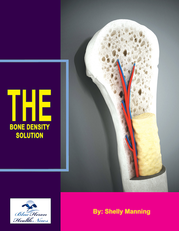
The Bone Density Solution by Shelly ManningThe program is all about healthy food and healthy habits. As we discussed earlier, we develop osteoporosis due to low bone density. Therefore, you will have to choose the right food to help your calcium and other vitamin deficiencies. In addition to healthy food, you will have to regularly practice some mild exercises. Your doctor might offer you the same suggestion. However, the difference is that The Bone Density Solution will help you with an in-depth guide.
Cortical vs. Trabecular Bone Density
Cortical and trabecular bones are the two primary types of bone tissue that differ in structure, function, and density. Understanding the differences in their density is essential for assessing overall bone strength and fracture risk. Here’s a detailed comparison between cortical bone density and trabecular bone density:
1. Cortical Bone (Compact Bone)
- Structure:
- Cortical bone forms the dense, outer layer of bones. It accounts for about 80% of the total bone mass in the human body.
- It is tightly packed, with minimal spaces, giving it a smooth, solid appearance. Cortical bone is responsible for the majority of the bone’s mechanical strength and rigidity.
- Density:
- High Density: Cortical bone is highly dense because it has fewer spaces between the bone cells. This compactness provides significant structural support, making it stronger and more resistant to bending and torsion forces.
- Slow Remodeling: Cortical bone remodels at a slower rate compared to trabecular bone. Its dense structure allows it to resist fractures under normal conditions, but when bone density decreases, cortical bone can weaken, particularly in areas like the hip and long bones.
- Location:
- Found primarily in the shafts of long bones (such as the femur and humerus), as well as in the outer surfaces of other bones.
- Role in Fracture Prevention:
- Cortical bone provides the rigidity and strength needed to support weight-bearing activities and protect internal organs. A decrease in cortical bone density, as seen in osteoporosis, increases the risk of fractures in long bones and hip fractures.
2. Trabecular Bone (Cancellous or Spongy Bone)
- Structure:
- Trabecular bone has a porous, lattice-like structure with many small, interconnected spaces. This spongy architecture allows it to absorb shock and distribute forces throughout the bone.
- Trabecular bone constitutes about 20% of bone mass but has a higher surface area compared to cortical bone, making it more metabolically active.
- Density:
- Lower Density: Trabecular bone is less dense than cortical bone because it contains more spaces and less compact bone tissue. This porous structure allows for flexibility and shock absorption, but it also makes trabecular bone more vulnerable to rapid bone loss and fractures.
- High Remodeling Rate: Trabecular bone undergoes more rapid remodeling than cortical bone. This high turnover rate makes it more responsive to changes in metabolic conditions, such as hormonal fluctuations or nutritional deficiencies. Loss of trabecular bone density occurs quickly in conditions like osteoporosis.
- Location:
- Found primarily in the ends of long bones, within the vertebrae of the spine, the pelvis, and other areas like the ribs.
- Role in Fracture Prevention:
- Trabecular bone helps distribute stress and absorb impacts, particularly in areas subjected to compressive forces like the spine and hip. Reduced trabecular bone density increases the risk of fractures in these regions, such as vertebral compression fractures or hip fractures.
3. Differences in Bone Density Between Cortical and Trabecular Bone
- Cortical Bone:
- Higher density due to its compact structure.
- Lower surface area, meaning it remodels more slowly and is less responsive to metabolic changes.
- Provides mechanical strength and resistance to bending and twisting forces.
- Loss of cortical bone density is more gradual but can lead to significant issues in weight-bearing bones like the femur and tibia.
- Trabecular Bone:
- Lower density due to its spongy structure.
- Higher surface area, meaning it remodels quickly and is more affected by hormonal and metabolic changes.
- Provides shock absorption and distribution of forces within the bone, particularly in the spine and pelvis.
- Loss of trabecular bone density can occur rapidly, increasing the risk of fractures, especially in areas like the spine and hips.
4. Impact of Aging on Cortical and Trabecular Bone Density
- Cortical Bone Loss:
- As people age, cortical bone density declines slowly. With aging, bone remodeling in cortical bone becomes less balanced, leading to thinning of the outer bone layer (cortical thinning). This loss is particularly notable in postmenopausal women due to hormonal changes.
- Decreased cortical bone density contributes to fractures in the long bones and hip fractures, which are more common in older adults.
- Trabecular Bone Loss:
- Trabecular bone density decreases more rapidly than cortical bone, particularly after menopause in women when estrogen levels drop. This accelerated loss makes trabecular bone more susceptible to fractures in areas like the spine and hip.
- Loss of trabecular bone density has a significant impact on fracture risk, particularly for vertebral compression fractures and hip fractures.
5. Role in Osteoporosis
- Cortical Bone and Osteoporosis:
- In osteoporosis, cortical bone thins and loses density, which compromises the bone’s ability to withstand mechanical forces. This leads to an increased risk of fractures, particularly in the hip and long bones.
- Trabecular Bone and Osteoporosis:
- Osteoporosis more significantly affects trabecular bone because of its higher remodeling rate. The porous structure becomes weaker, leading to fractures in the spine and other regions where trabecular bone is predominant.
6. Measurement of Bone Density
- Cortical Bone Density: Measured using techniques like DEXA (Dual-Energy X-ray Absorptiometry) scans, typically focusing on areas like the femur (thigh bone) and forearm to assess cortical bone mass.
- Trabecular Bone Density: DEXA scans and other imaging techniques (like Quantitative Computed Tomography or QCT) are used to assess trabecular bone, often focusing on the spine, hip, or pelvis, where trabecular bone is abundant.
Summary:
- Cortical Bone Density: High density, provides structural strength and support, remodels slowly, and is crucial for resistance to bending and weight-bearing forces.
- Trabecular Bone Density: Lower density, provides shock absorption and distributes mechanical stress, remodels rapidly, and is more susceptible to bone loss and fractures due to hormonal or metabolic changes.
Both cortical and trabecular bones play essential roles in maintaining overall bone strength and function. The balance between the two types of bone tissue—and how their densities change over time—significantly affects the risk of fractures, especially in conditions like osteoporosis.
The Bone Density Solution by Shelly ManningThe program is all about healthy food and healthy habits. As we discussed earlier, we develop osteoporosis due to low bone density. Therefore, you will have to choose the right food to help your calcium and other vitamin deficiencies. In addition to healthy food, you will have to regularly practice some mild exercises. Your doctor might offer you the same suggestion. However, the difference is that The Bone Density Solution will help you with an in-depth guide.